
Figure 2. Apoptosis induced by asparaginase is partially caspase 3-dependent in K562 CML cells.
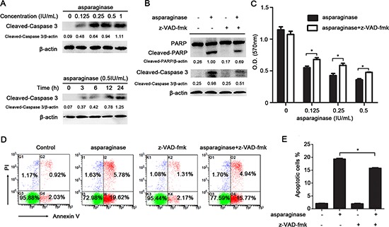
(A) K562 cells were dose- and time-dependently incubated with asparaginase, then western blot analysis was performed to assess the level of cleaved-caspase 3. Densitometric values were quantified using the ImageJ software, and the data represented mean of three independent experiments. (B) K562 cells were incubated with 0.5 IU/mL of asparaginase, either alone or in combination with 20 μM z-VAD-fmk for 24 h, then western blot analysis was performed to assess the level of cleaved-caspase 3, PARP and cleaved-PARP. Densitometric values were quantified using the ImageJ software, and the data are presented as means ± SD of three independent experiments. (C–E) K562 cells were treated with asparaginase at indicated concentrations in the absence or presence of 20 μM z-VAD-fmk for 48 h. (C) Cell viability was determined by MTT assay at the wavelength of 570 nm. (D) Cells were stained with Annexin V/PI and analyzed by flow cytometry after 48 h incubation. (E) The percentages of Annexin V-positive/PI-negative cells were presented in bar charts. Results were represented as mean ± SD (*P < 0.05).