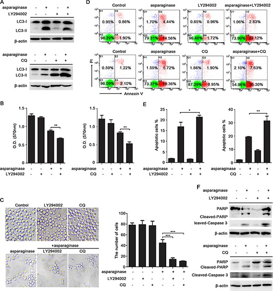
Figure 4. Inhibition of autophagy enhances asparaginase-induced K562 cell death.

(A) K562 cells were treated with 0.04 IU/mL of asparaginase in the absence or presence of 20 μM LY294002 or 10 μM CQ for 24 h, autophagy-associated protein LC3-I/II were detected by western blot analysis. (B–E) K562 cells were incubated with 0.04 IU/mL of asparaginase in the absence or presence of 20 μM LY294002 or 10 μM CQ for 48 h. (B) Cell viability was analyzed by MTT assay. (C) Morphological and numerary changes of K562 cells were observed using microscopy and photography. The number of normal cells was presented in bar charts. (D) Cell apoptosis was detected by Annexin V-FITC/PI staining. (E) The percentage of Annexin V-positive/PI-negative K562 cells was presented in bar charts. (F) K562 cells were treated with 0.04 IU/mL of asparaginase in combination with or without 20 μM LY294002 or 10 μM CQ for 24 h, the expression level of protein cleaved-caspase 3, PARP and cleaved-PARP were analyzed by western blot analysis. Results were represented as mean ± SD (*P < 0.05, **P < 0.01, ***P < 0.001).
