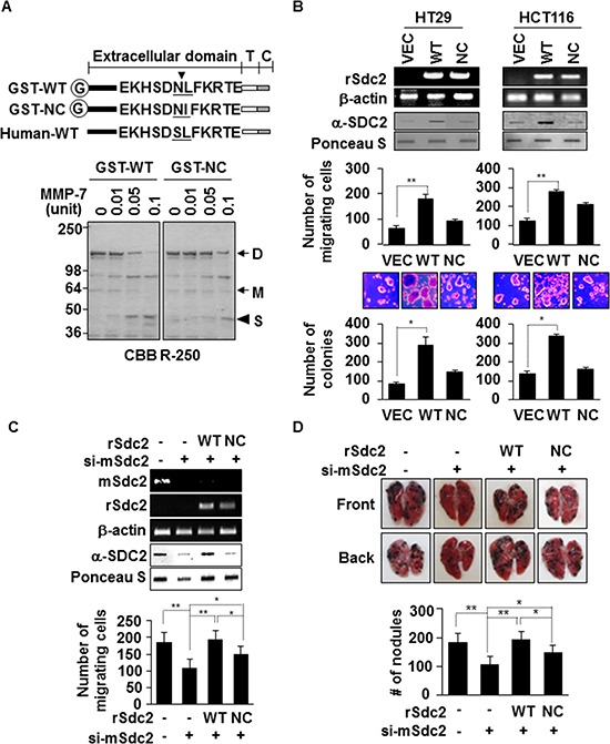
Figure 1. Extracellular domain shedding is necessary for syndecan-2 functions in colon cancer.

(A) Equivalent amounts of GST-fused rat syndecan-2 wild-type (WT) or the Leu149® Ile non-cleavable mutant (NC) were incubated with pro-MMP-7, separated by SDS-PAGE, and stained with Coomassie Brilliant Blue. The arrows indicate the dimeric (D) and monomeric (M) forms of syndecan-2. The arrowhead indicates the shed fragment of syndecan-2 (S). (B) Colon cancer cells were transfected with 1 μg of VEC, WT- or NC-syndecan-2. Syndecan-2 mRNA expression was evaluated by RT-PCR using the indicated primer. Conditioned medium was subjected to slot blotting with the anti-syndecan-2 antibody (top). Transfected cells were allowed to migrate on gelatin-coated (10 μg/ml) Transwell plates, and migrated cells were stained with hematoxylin and eosin, and counted (middle). n = 3; **p < 0.01. Transfected cells were added to bottom agar and colonies were counted after 2 weeks (bottom). n = 5; *p < 0.05. (C) B16F10 cells were transfected with siRNA targeting mouse syndecan-2 alone or with 1 μg of the indicated rat syndecan-2 constructs. Syndecan-2 mRNA expression, shed syndecan-2 in the conditioned media and cell migration were analyzed as described in Figure 1B. n = 5; **p < 0.01, *p < 0.05. (D) BALB/c mice were injected with the indicated B16F10 melanoma cells. After 2 weeks, lungs were removed and examined. Two independent experiments were performed (n = 5/each group). Representative photographs of the front and back sides of each lung are shown (top). The bar graph indicates the numbers of metastatic lung nodules (bottom). The columns represent the mean (± s.d.) number of lung metastatic nodules (n = 3). *p < 0.05, **p < 0.01.
