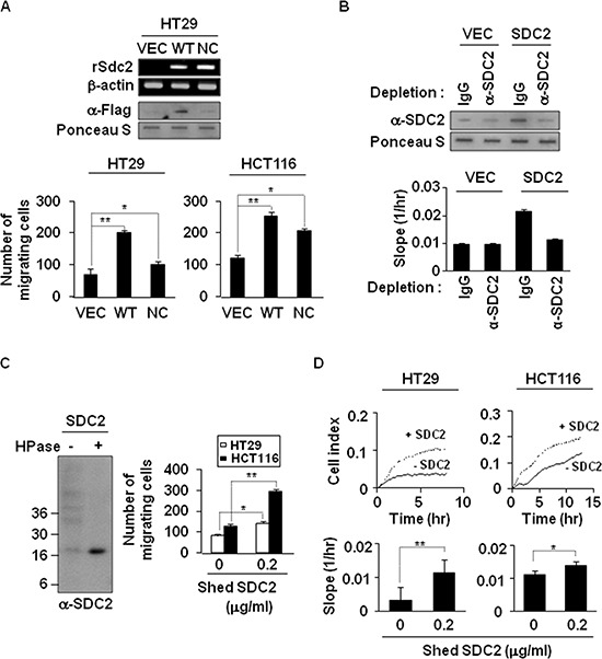
Figure 2. Shedding of syndecan-2 plays a critical role in colon cancer cell migration.

(A) HT29 cells were transfected with indicated cDNAs, and syndecan-2 mRNA expression was evaluated by RT-PCR. Conditioned media were subjected to slot blotting with the anti-Flag antibody (top). HT29 and HCT116 cells were treated with HT29 conditioned media (final 10% v/v) from VEC, WT-or NC-syndecan-2 mutant transfected cells and allowed to migrate on Transwell apparatus (bottom). n = 5; *p < 0.05, **p < 0.01. (B) Conditioned media were immunodepleted with control IgG- or anti-syndecan-2 antibody-conjugated protein G beads. The supernatants were subjected to slot blotting with anti-syndecan-2 antibody (top). Mixture of HCT116 cells with each supernatant (final 10% v/v) were added to the upper chambers of CIM-plates and migration curves were monitored using the xCELLigence system. The rates of cell migration over 24 hr were analyzed using the RTCA software to each RTCA CIM-Plate wells (bottom). (C) Shed syndecan-2 in HT29 cell conditioned media was isolated by DEAE-Sepharose column chromatography. Final elution fractions were digested by heparinase and analyzed by immunoblotting using anti-syndecan-2 antibody (left). Cells were treated with 0.2 μg/ml of purified shed syndecan-2, and Transwell migration assay was performed (right). n = 5; *p = 0.05, **p < 0.01. (D) Cells were treated with 0.2 μg/ml of purified shed syndecan-2, and a real-time migration assay was analyzed by xCELLigence system. n = 5; *p = 0.05, **p < 0.01.
