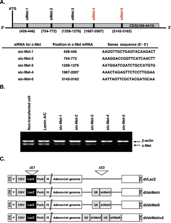
Figure 1. Schematic and characterization of c-Met RNAi target site.

(A) Location of five c-Met-specific siRNAs examined in this study. The target sequences within c-Met are shown. (B) shRNA-mediated in vitro knockdown of c-Met gene. Cells were transfected for 48 hr with pSP72/U6-sic-Met1, pSP72/U6-sic-Met2, pSP72/U6-sic-Met3, pSP72/U6-sic-Met4, or pSP72/U6-sic-Met5. LaminA/C was used as negative control. The knockdown of endogenous expression was measured by reverse transcriptase-polymerase chain reaction (RT-PCR) for c-Met. The experiment was repeated three times with reproducible results. (C) Schematic representation of the genomic structures of dl/LacZ, dl/shMet4, dl/shMet5, and dl/shMet4+5 adenoviruses used in this study.
