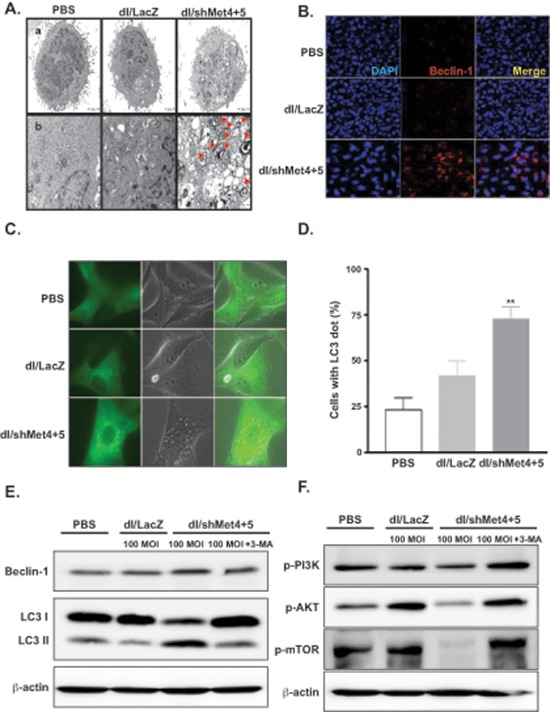
Figure 4. Induction of autophagy in glioma cells transduced with c-Met-specific shRNA-expressing Ad.

(A) Electron photomicrographs showing the ultrastructure of U343 cells transduced with dl/LacZ or dl/shMet4+5 at an MOI of 50 for 72 hr. The arrowheads indicate autophagic vacuoles. Scale bar = 2 μm. Original magnification: × 5000 (top) and × 10000 (bottom). (B) Microscope image of Beclin-1. U343 cells were transduced with dl/LacZ or dl/shMet4+5 at an MOI of 30 for 72 hr. Cells were then labeled with anti-Beclin-1 (red) or DAPI (blue), and examined by confocal microscopy. Original magnification: × 400. (C) U87MG cells stably transfected with GFP-LC3 were transduced with dl/LacZ or dl/shMet4+5 at an MOI of 100 for 48 hr. And then the translocation of GFP-LC3 was observed. Representative images of the cells were observed under fluorescence and phase-contrast microscopy. Original magnification: × 600. (D) The percentage of cells with GFP-LC3 localized to granular structures was estimated by counting a minimum of 100 cells per sample, and data is represented as means ± SE of five independent experiments. **P < 0.01 compared with PBS-treated and dl/LacZ-transduced cells. (E) Involvement of LC3 and Beclin-1 in shMet-expressing Ad-transduced glioma cells. U343 cells were pretreated with or without 3-MA (0.5 mM) for 30 min, and then transduced with dl/LacZ or dl/shMet4+5 at 100 MOI. Cells were then analyzed with antibody against Beclin-1 or LC3-I & -II (microtubule associated protein 1 light chain 3). (F) Suppression of c-Met-induced PI3K–AKT–mTOR signaling pathway. U343 cells were treated as indicated above (Figure 4E). After 48 hr, cell lysates were determined by western blot analysis with antibodies against p-PI3K, p-AKT, and p-mTOR.
