
Figure 4. The involvement of miR-191 in the 5-Fu-induced cell apoptotic pathway in HCT116 cells.
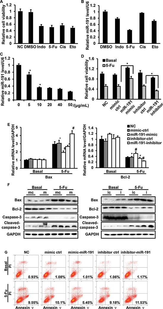
HCT116 cells were treated with various commonly-used chemotherapeutic drugs, including 5-fluorouracil (5-Fu, 10 μg/ml), indomethacin (Indo, 10 μg/ml), cisplatin (Cis, 10 μg/ml) and etoposide (Eto, 2 μM). (A) Cell viability was assessed by CCK8 assays. (B) RT-PCR analysis of the relative expression of miR-191. (C) 5-Fu down-regulated miR-191 in a dose-dependent manner. HCT116 cells were treated with the indicated concentration of 5-Fu for 48 hours, total mRNA was isolated, and the miR-191 level was analyzed by RT-PCR (*P < 0.05 versus DMSO treated cells). HCT116 cells were transfected with the indicated oligos for 24 hours and then treated with 5-Fu (25 μg/ml) for another 24 hours. (D) Cell viability was assessed by CCK8 assays (*P < 0.05 versus 5-Fu treated mimic-ctrl/inhibitor-ctrl cells). (E) The mRNA levels of Bax and Bcl-2 was determined by RT-PCR (*P < 0.05 versus DMSO treated mimic-ctrl/inhibitor-ctrl cells, #P < 0.05 versus 5-Fu treated mimic-ctrl/inhibitor-ctrl cells). (F) The protein levels of Bax, Bcl-2, caspase-3 and cleaved-caspase-3 were detected by western blotting analysis in HCT116 cells transfected with mimic-ctrl/miR-191-mimic (left panel) and inhibitor-ctrl/miR-191-inhibitor (right panel) after DMSO or 5-Fu treatment. (mc, mimic ctrl; m, miR-191-mimic; ic, inhibitor ctrl; i, miR-191-inhibitor) (G) Cell apoptosis analysis of transfected cells after treatment. GAPDH served as a loading control. The data represents the means ± SDs.