
Figure 2. WISP-1 regulates the angiogenesis by raising VEGF-A expression in OSCC cells.
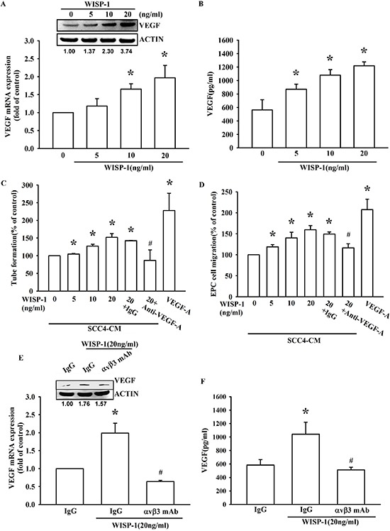
(A–B) SCC4 cells were incubated with WISP-1 (0–20 ng/mL) for 24 h, VEGF-A expression was measured by qPCR, ELISA, and western blot. (C–D) SCC4 cells were incubated with WISP-1 (0–20 ng/mL) for 24 h, and the CM was collected. EPCs were pre-treated for 30 min with IgG control antibody or VEGF-A antibody (1 μg/mL) and incubated with CM for 6 h and cell capillary-like structure formation in EPCs was examined by tube formation assay (C) EPCs were incubated with CM for 24 h, and cell migration was examined using the transwell assay (D) (E–F) SCC4 cells were incubated with the integrin αvβ3 antibody for 30 min, followed by stimulation with WISP-1 (20 ng/mL) for 24 h. VEGF-A expression was examined by western blot, qPCR, and ELISA. Data are expressed as mean ± SEM *P < 0.05 compared to control; #P < 0.05 compared to the WISP-1 treated group.