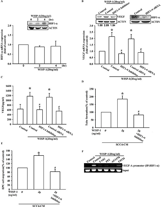
Figure 5. WISP-1 promotes VEGF-A expression in OSCC and contributing to angiogenesis through the HIF1-α signaling pathway.

(A) SCC4 cells were stimulated by WISP-1 (20 ng/mL) for the indicated times (0, 2, and 4 h). HIF1-α expression level was measured by western blot and qPCR. (B–E) SCC4 cells were pre-treated with HIF1-α inhibitor (1 μM) for 30 min or transfected with HIF1-α siRNAs for 24 h, followed by WISP-1 (20 ng/mL) stimulation for 24 h. The assay procedures were performed as described in Figure 3A–3D. (F) SCC4 cells were incubated with FAKi, PP2, AG1478, or U0126 for 30 min, followed by stimulation with WISP-1 (20 ng/mL) for 60 min. Chromatin immunoprecipitation (ChIP) assays were performed using an anti-HIF1-α antibody. One percent of the precipitated chromatin was analyzed to verify equal loading (input). Data are expressed as the mean ± SEM. *P < 0.05 compared with control; #P < 0.05 compared with the WISP-1-treated group.
