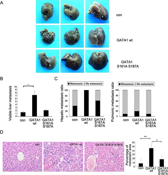
Figure 5. Phosphorylation of GATA1 enhances breast cancer cell metastasis in vivo.

1 × 10⁷ MCF-7 cells stably expressing empty vector, GATA1 wt or GATA1 S161A S187A were injected into nude mice through tail vein. (A) Livers were dissected after injection and macroscopically photographed. (B) Graphical representation of the number of liver metastases from each mouse (mean ± s.d.); *p < 0.05. (C) Effect of MCF7 cells with stable expression of control Flag, Flag GATA1 wt, Flag GATA1 S161A S187A on incidence of liver and lung metastasis. (D) Representative image of H&E-stained liver sections of mice and percentage of metastatic liver surface area relative to tatal live surface area. scale bar, 50 μm. 30 independent visions were counted. *p < 0.05, **p < 0.01.
