
Figure 6. The phosphorylated GATA1 recruits more HDAC3/4 to promote transcriptional repression of E-cadherin.
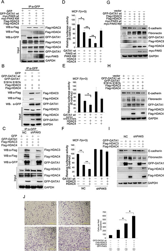
(A) HEK-293 cells were transfected with GFP-GATA1, Myc-PAK5wt/KM, Flag-HDAC3/4 as indicated. Lysates were immunoprecipitated with anti-GFP antibody and immunoblotted with anti-Flag, anti-GFP and anti-myc antibody. (B) HEK-293 cells were co-transfected with different combination of plasmids as indicated. Lysates were immunoprecipitated with anti-GFP antibody and immunoblotted with anti-Flag and anti-GFP antibody. (C) MCF-7 cells stably infected with shPAK5 were co-transfected with different combination of plasmids as indicated. Lysates were immunoprecipitated with anti-GFP antibody and immunoblotted with anti-Flag and anti-GFP antibody. (D) PAK5wt/KM and GATA1, HDAC3/4 were transfected into MCF-7 cells as indicated for Luciferase Assays. *p < 0.05, **p < 0.01. (E) GATA1 wt and GATA1 S161A S187A, HDAC3/4 were transfected into MCF-7 cells as indicated for Luciferase Assays. *p < 0.05. (F) GATA1, HDAC3/4 were transfected into stably PAK5 knockdown MCF-7 cells as indicated for Luciferase Assays. **p < 0.01. (G–I) The same transfection as D, E, F cell lysates from these cells were used for immunoblotting using anti-Fibronectin, E-cadherin antibodies. (J) GATA1, HDAC3/4 and PAK5 were transfected into MCF-7 cells, then transwell assay was used. Results are representative of three independent experiments. Migrated cells were plotted as the average number of cells per field of view. *p < 0.05.