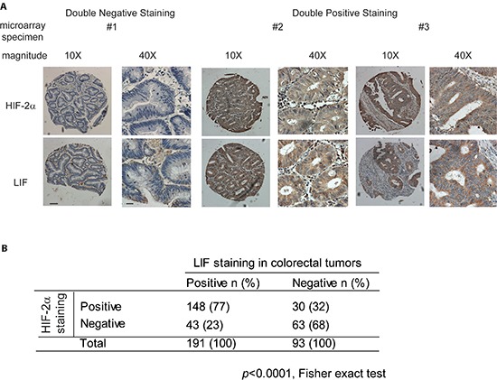
Figure 5. HIF-2α overexpression is associated with LIF overexpression in human colorectal cancer specimens.

HIF-2α and LIF protein levels were determined by IHC staining in tissue microarrays containing 284 cases of human colorectal cancer specimens. (A) Representative IHC staining results for HIF-2α (upper panels) and LIF (lower panels) are shown. Positive HIF-2α or LIF staining: > 10% cells stained with HIF-2α or LIF, respectively. Scale bar: 50 μm for low magnitude images (10X); 10 μm for high magnitude images (40X). (B) HIF-2α overexpression is associated with LIF overexpression in human colorectal tumors (p < 0.0001, Fisher exact test).
