Figure 7.
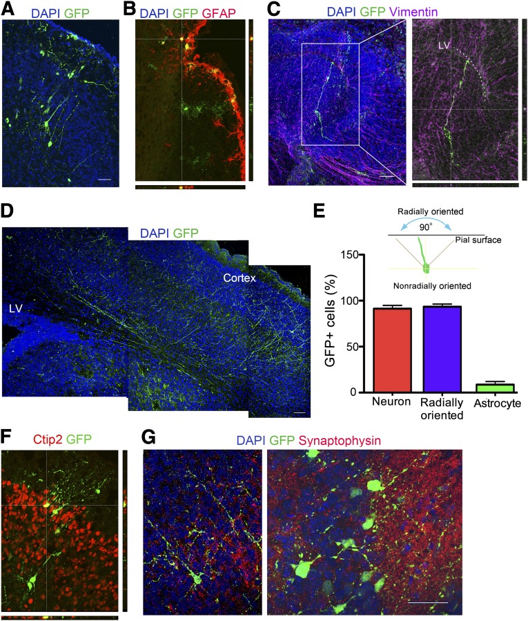
Differentiation, migration, and integration of engrafted GFP-labeled human embryonic stem cell-derived radial glia (RG) in mouse brain. E14 mouse brains were injected, and the brains were examined on postnatal day 3. (A): GFP-positive pyramidal neurons were radially arranged in the cortex. (B): GFP-positive cells with astrocyte morphology colocalize with GFAP in the outermost lining of the cortex. (C): Bipolar GFP-expressing cells with a RG-like morphology localize to the P3 LV, parallel to host vimentin-positive radial cells. Magnification, ×20 (left), ×40 (right). (D): Extensive GFP-positive radially oriented processes start near the LV and extend to the cortex. (E): Radial orientation is defined as the direction of neurites in the symmetrical 90° region perpendicular to the pial surface. Quantification of cells with neuronal or astrocytic morphology among the total GFP positive cells is shown. The percentage of radially oriented cells among the cells with neuronal morphology is also indicated. Data are mean ± SEM, n = 9. (F): GFP-positive neurons localize to the correct cortical layer and express the layer-specific marker Ctip2. (G): Dotted synaptophysin stained host processes make contact with GFP-positive neuronal processes. Magnification, ×20 (left), ×63 (right). Scale bars = 50 μm. Abbreviations: DAPI, 4′,6-diamidino-2-phenylindole; GAFP, glial fibrillary acidic protein; GFP, green fluorescent protein; LV, lateral ventricle; P, passage.

