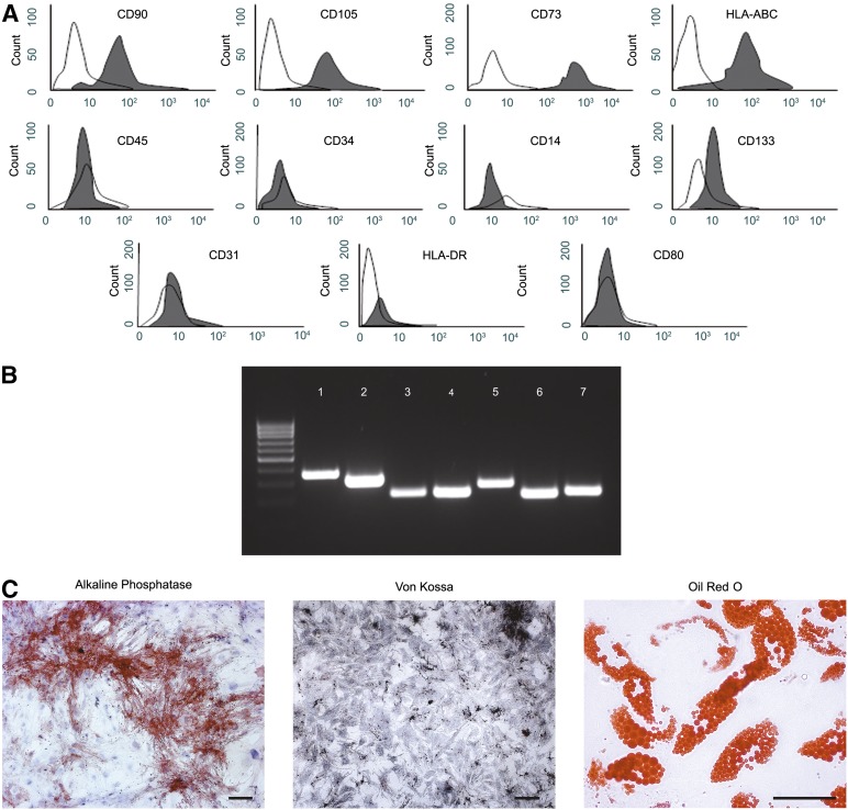
Figure 1.
Characterization of human amniotic membrane-derived mesenchymal stromal cells (hAMCs). (A): Fluorescence-activated cell sorting analysis of hAMCs at passage 3. (B): Reverse transcription-polymerase chain reaction analysis for osteocyte markers osteopontin (band 1), cathepsin K (band 2), and bone sialoprotein (band 3) or adipocyte markers peroxisome proliferation-activated receptor γ (band 5) and adipose differentiation-related protein (band 6), after an osteogenic or adipogenic differentiation assay. Glyceraldehyde 6-phosphate dehydrogenase (bands 4 and 7) was used as an endogenous control. (C): Cytochemical analysis: alkaline phosphatase activity assay (bright field, scale bar = 100 µm) and Von Kossa staining (bright field, scale bar = 100 µm) of hAMCs after osteogenic induction and Oil Red O staining (bright field, scale bar = 50 µm) of hAMCs at the end of the adipogenic differentiation assay. Data are representative of three independent experiments. Abbreviation: HLA, human leukocyte antigen.

