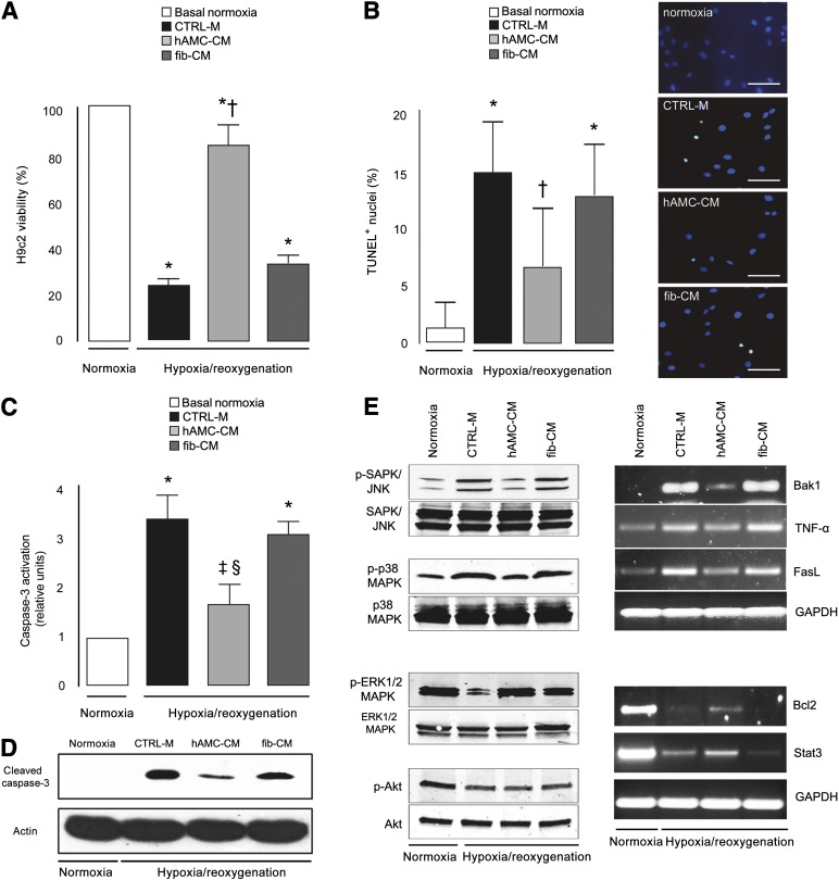
Figure 2.
Cytoprotective effect of hAMC-CM in vitro. (A): MTS viability assay. (B): Quantitative analysis and representative images of the TUNEL assay. Nuclei are shown in blue, and TUNEL+ in green. (C): Colorimetric assay of caspase-3 activity. (D): Immunoblotting for cleaved caspase-3 (17 kDa). (E): Representative images of Western blot (left) and reverse transcription-polymerase chain reaction (right) analysis. ∗, p < .001 versus basal normoxia; †, p < .001 versus CTRL-M and fib-CM; ‡, p < .001 versus CTRL-M; §, p < .01 versus fib-CM. All data expressed as the mean ± SD. Data are representative of three independent experiments. Abbreviations: Bak1, Bcl-2 antagonist/killer 1; Bcl-2, B-cell lymphoma 2; CTRL-M, control medium; ERK1/2 MAPK, extracellular signal-regulated kinase 1/2 mitogen-activated protein kinase; FasL, Fas ligand; fib-CM, human dermal fibroblast conditioned medium; GAPDH, glyceraldehyde 6-phosphate dehydrogenase; hAMC-CM, conditioned medium obtained from human amniotic membrane-derived mesenchymal stromal cells; p, phosphorylated; p38 MAPK = p38 mitogen-activated protein kinase; SAPK/JNK, stress-activated protein kinase/Jun amino-terminal kinase; Stat3, signal transducer and activator of transcription 3; TNF-α, tumor necrosis factor-α; TUNEL, terminal deoxynucleotidyl transferase dUTP nick-end labeling.

