Figure 3.
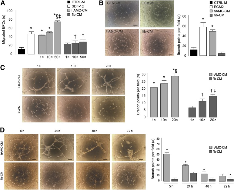
hAMC-CM promotes angiogenesis in vitro. (A): Boyden chamber migration assay. ∗, p < .01 versus CTRL-M and fib-CM 1×, 10×, and 50×; †, p < .001 versus CTRL-M; §, p < .01 versus SDF-1α; ‡, p < .01 versus hAMC-CM 1× and 10×. (B): Representative images of EPCs cultured in Matrigel for 5 hours in the presence of CTRL-M, EGM2 (growth medium), hAMC-CM, or fib-CM and quantification of the number of branches per field. Scale bar = 200 µm. ∗, p < .001 versus CTRL-M and fib-CM. (C): Representative images of EPCs cultured in Matrigel for 24 hours in the presence of hAMC-CM or fib-CM at 1×, 10×, or 20×. Bars summarize the quantitative analysis. Scale bar = 200 µm. ∗, p < .001 versus fib-CM 1×, 10×, and 20×; §, p < .001 versus hAMC-CM 1× and 10×; †, p < .05 versus fib-CM 1×; ‡, p < .05 versus fib-CM 10×. (D): Representative images of EPCs cultured in Matrigel in the presence of hAMC-CM 20× or fib-CM 20× for 5, 24, 48, and 72 hours; bars represent the quantitative analysis. Scale bar = 200 µm. ∗, p < .001 versus fib-CM. All data are expressed as the mean ± SD. Data are representative of three independent experiments. Abbreviations: CTRL-M, control medium; EGM2, endothelial growth medium 2; EPCs, endothelial progenitor cells; fib-CM, human dermal fibroblast conditioned medium; hAMC-CM, conditioned medium obtained from human amniotic membrane-derived mesenchymal stromal cells; SDF-1α, stromal cell derived factor-1α.

