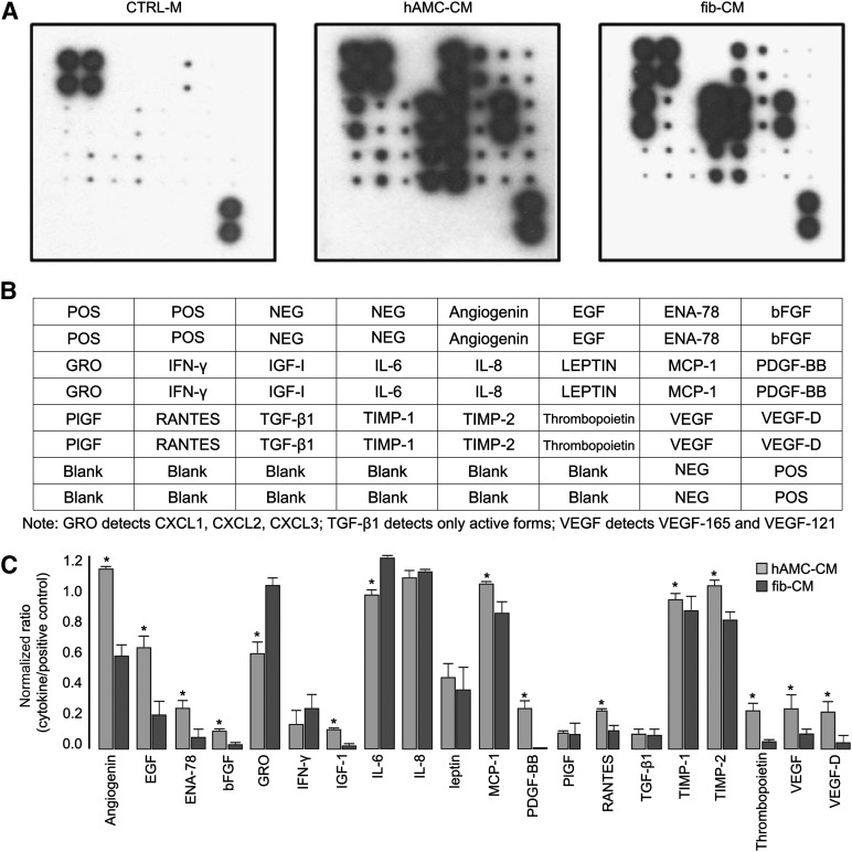
Figure 7.
Proangiogenic factors present in the hAMC-CM. (A): Representative images of human cytokine antibody array membranes (RayBio Human Angiogenesis Antibody array I, RayBiotech, Norcross, GA, http://www.raybiotech.com) of CTRL-M, hAMC-CM, and fib-CM. (B): Layout of the membrane for the human cytokine antibody array. Positive controls are located in the upper left corner (four spots) and lower right corner (two spots) of each membrane. (C): Quantitative analysis confirmed that several proangiogenic cytokines are significantly more abundant in hAMC-CM than in fib-CM. ∗, p < .05 versus fib-CM. All data are expressed as the mean ± SD. Data are representative of two independent experiments. Abbreviations: bFGF, basic fibroblast growth factor; CTRL-M, control medium; EGF, endothelial growth factor; ENA-78 or CXCL5, C-X-C motif chemokine 5; fib-CM, human dermal fibroblast conditioned medium; GRO, growth-regulated oncogene; hAMC-CM, human amniotic membrane-derived mesenchymal stromal cells conditioned medium; IFN-γ, interferon-γ; IGF-1, insulin-like growth factor 1; IL, interleukin; MCP-1, monocyte chemoattractant protein-1; Neg, negative; PIGF, placental growth factor; PDGF, platelet-derived growth factor; Pos, positive; RANTES, regulated on activation, normal T cell expressed and secreted; TGF-β1, transforming growth factor-β1; TIMP, tissue inhibitor of metalloproteinase; VEGF, vascular endothelial growth factor.

