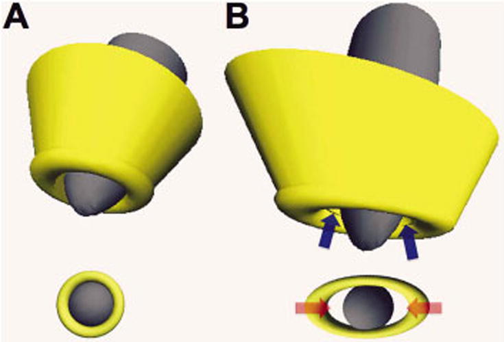
Fig. 5.

Three-dimensional reconstruction of the root apical region and shape of the physiological foramen. In A, a root with a conical canal can be seen that ends in a round physiological foramen constructed with the values of maximum and minimum diameters obtained from the MV25 sample, where when the positioning of a round endodontic instrument or one that generates a round preparation is simulated, it adapts completely to the physiological foramen. In B, a root with more parallel canal walls that end at an oval-shaped physiological foramen with the maximum and minimum diameter values obtained from the D35 sample, where when the positioning of a round endodontic instrument or one that generates a round preparation is simulated, it is only adapted to the most central area of the foramen, a limit due to its minimum diameter, leaving areas of the physiological foramen (blue arrows) uninstrumented, and spaces that can house remains of tissue and microorganisms (red arrows) that will then be able to access the periapical tissues.
