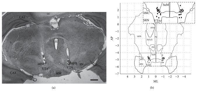
Figure 7.
Histological assessment. Electrode placement was assessed using H&E staining of brain slices (a), with the bilateral positions of individual electrode tips summarized (b). Electrode tips were within the dorsoventral range of −8.0 ± 0.1 mm, according to Paxinos Rat Brain Atlas [12]. CA1: CA1 field of the hippocampus; CA3: CA3 field of the hippocampus; MPA: medial preoptic area; mfb: medial forebrain bundle; PeFLH: perifornical part of lateral hypothalamus; AHA: anterior hypothalamic area; PH: posterior hypothalamus; 3V (dorsal): third ventricle; MM: medial mammillary nucleus; cc: corpus callosum; SuM, supramammillary nucleus; VTA: ventral tegmental area; SNC: substantia nigra pars compacta; SRN: substantia nigra pars reticulate; AP: anterior-posterior; ML: mediolateral. Scale bar = 500 µm.

