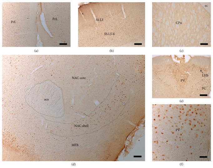
Figure 8.
Immunohistochemical visualization of c-fos expression. Following MFB-HFS, strong c-fos expression was observed in prelimbic cortex (a), layers 3 and 5/6 of the somatosensory cortex (b), paraventricular thalamus, lateral habenula (e, f), and shell of the nucleus accumbens (d), suggesting that the stimulation activated mesolimbic and mesocortical pathways and its projection targets. However, little or no c-fos expression was observed in the dorsal striatum where the stimulated pathways do not project to. See Table 1 and text for more details. PrL: prelimbic cortex; NAC: nucleus accumbens; PV: paraventricular thalamic nucleus; LHb: lateral habenular nucleus; PC: paracentral thalamic nucleus; aca: anterior commissure; SS: somatosensory cortex; ec: external capsule; CPu: striatum. Scale bar = 50 µm in (a), (b), (d), and (e); 250 µm in (c) and (f).

