Fig. 7.

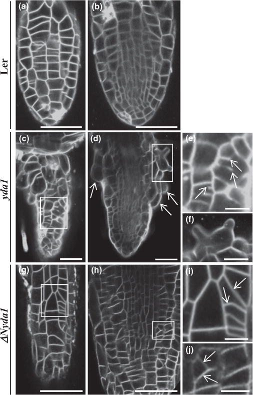
Cell division patterns as visualized in young emerging lateral roots of Arabidopsis Landsberg erecta (Ler), yda1 and ΔNyda1 stained with membrane marker FM4-64 and imaged with CLSM. (a, b) Epidermal (a) and central (b) optical sections of a lateral root of Ler showing the fairly ordered cell arrangement of epidermal and central lateral root tissues. (c, d) Epidermal (c) and central (d) optical sections of a yda1 lateral root exhibiting abnormal cell patterning (c) and bulging of epidermal cells (d, arrows). (e) Higher magnification of the outlined area in (c) showing incomplete cell plate formation in two cells (arrows). (f) Higher magnification of the outlined area in (d) showing a radially expanded trichoblast with two emerging bulges. (g, h) Epidermal (g) and central (h) optical sections of the same ΔNyda1 lateral root showing variably disturbed cell division planes (outlined areas). (i, j) Magnified views of the respective outlined areas in (g) and (h) showing oblique cell plates (arrows). Bars: (a–d, g, h) 50 μm; (e, f, i, j) 10 μm.
