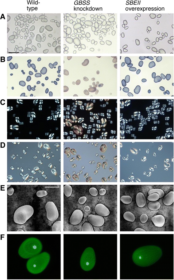
Figure 5.

Images of tuber starch granules from wild-type and transgenically modified potato tubers. (A) Brightfield. (B) Brightfield, starch stained with I2/KI. (C) Polarised light. (D) Differential interference contrast. (E) Variable pressure scanning electron microscopy. (F) Optical section of starch fluorescently labelled with APTS taken using confocal scanning laser microscopy. Lines used were WT898, 1041–3 and 1047–17. The scale bar (in panel A) for light microscopy pictures (A–D) represents 100 μm, and for the scanning electron microscopy (E) represents 40 μm.
