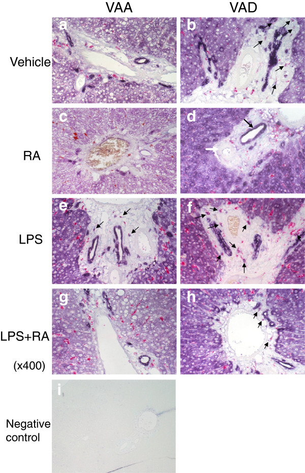
Figure 4.

Co-localization of ALDH1A1 protein expression and rat macrophage marker ED1 in liver from VAA and VAD rats. Dual IHC with anti-ALDH1A1 antibody and anti-ED1 antibody was performed followed by methyl green counterstaining for detection of nuclei. Anti-ALDH1A1 staining (purple) showed a broad distribution, which was intense around portal regions and bile ducts, while cells staining for ED1 (pink-red) was more generally scattered in the parenchyma and in portal areas after treatment with LPS (Figure 4 e and f compared with a and b). Magnification: x400. Panel i shows negative staining control.
