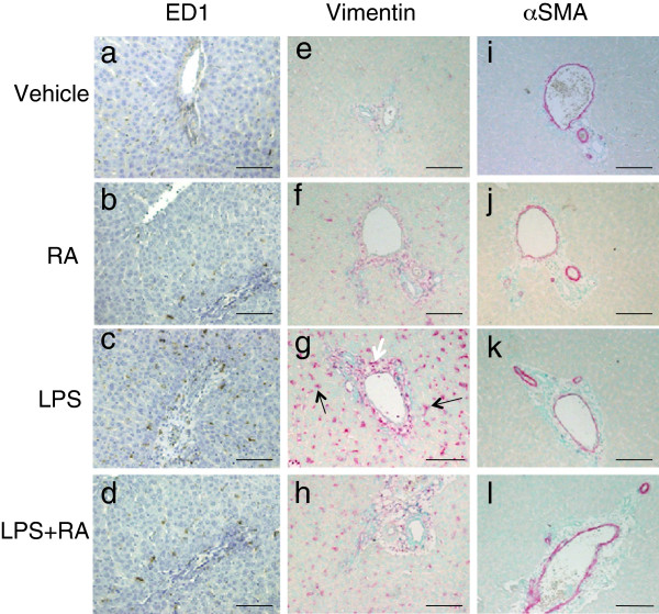
Figure 6.

Detection of macrophages (a-d), vimentin (e-h), and α-SMA (i-l) by IHC in liver of VAD rats. ED1 staining (brown) detected macrophages; vimentin (red) HSC/fibroblasts, and α-SMA (purple) cells containing smooth-muscle fibers. Nuclei were detected with methyl green dye. Numerous vimentin-positive star-shaped cells are present scattered in the parenchymal after treatment with LPS (panel g, black arrows) or LPS + RA (panel h). The white arrow in Figure 6g indicates an example of vimentin-positive cells located in the subendothelial mesenchyme. Scale bars; 100 μm.
