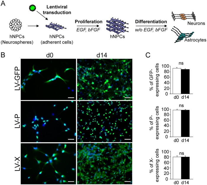
Fig 1. Lentiviral transduction and establishment of transgenic hNPCs.
(A) Schematic representation of the experimental procedure. (B) Immunofluorescence labeling of undifferentiated (day 0) and differentiated (day 14) hNPCs, following lentiviral transduction. Antibodies against the viral P (green) or X (green) proteins were used and nuclei were stained with DAPI (blue). Note the localization of the P (nuclear) and the X (nuclear and cytoplasmic) proteins. (C) Evaluation of transduction efficiency based on enumeration of immunostained cells. Results are representative of 3 independent experiments performed in triplicate. Statistical analyses were performed using the Mann-Whitney test. ns, non-significant (p > 0.5). Scale bar, 20 μm.

