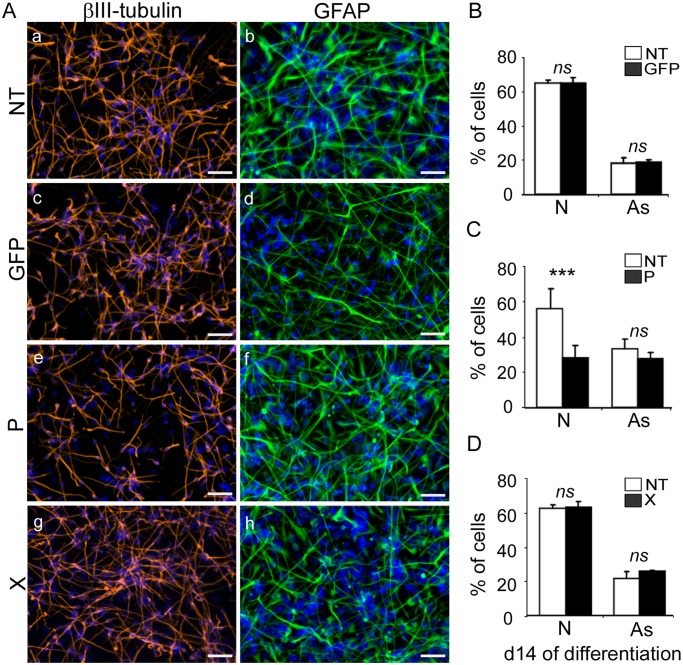
Fig 3. Expression of the bdv-p but not bdv-x gene in hNPCs impairs neuronal differentiation.
Transduced hNPCs expressing gfp, bdv-p or bdv-x genes and their matched NT controls were induced to differentiate for 14 days. (A) Immunostaining with antibodies directed against βIII-Tubulin, a neuronal marker (red), or GFAP, an astrocytic marker (green). Nuclei were stained with DAPI (blue). For panel homogenization, GFAP immunostaining performed in gfp-expressing hNPCs was re-colored in green. The percentage of neurons and astrocytes was determined based on enumeration of βIII-Tubulin-, GFAP- and DAPI-positive cells in (B) gfp-expressing hNPCs, (C) bdv-p-expressing hNPCs and (D) bdv-x-expressing hNPCs. Results in B, C and D are representative of 2, 5 and 2 independent experiments, respectively. All experiments were performed in triplicate. Statistical analyses were performed using the Mann-Whitney test. ***, p < 0.001, ns, non-significant (p > 0.5). N, neurons. As, astrocytes. Scale bar, 50 μm.

