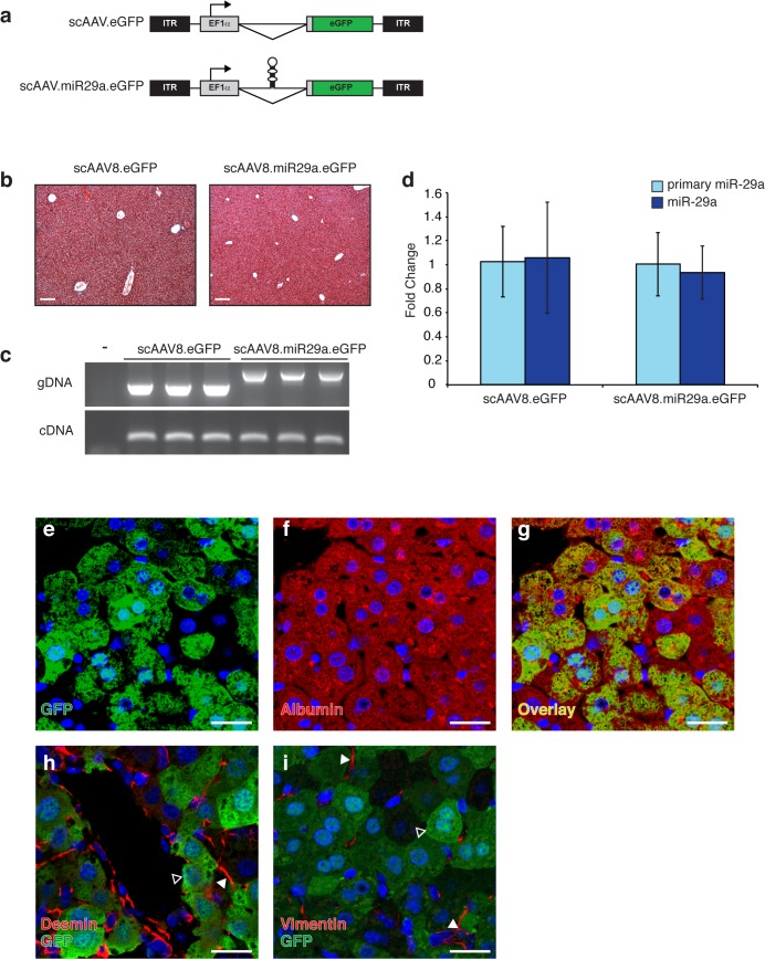
Fig 2. scAAV8 transduction and miR-29 expression levels in murine liver.
(a) Schematic representation of scAAV vectors depicting locations of inverted terminal repeats (ITRs), elongation factor 1-alpha promoter (EF1α), miRNA (shown in hairpin form), and enhanced green fluorescent protein (eGFP) open reading frame. (b) Transduction with scAAV8 does not disrupt normal liver architecture. Trichrome stained liver sections from AAV-transduced animals demonstrating normal histology. Scale bar = 100μm (c) Viral genomic DNA (gDNA) and mRNA from the EF1α transcription unit (cDNA) are readily detectable in mouse liver following transduction with 2x10¹¹ vg of scAAV8.eGFP (n = 3) or scAAV8.miR29a.eGFP (n = 3). The presence of the hairpin accounts for the increased size of the scAAV8.miR29a.eGFP gDNA amplicon. (d) Hepatic expression of primary and mature miR-29a in scAAV8 transduced mice. Average fold change for each treatment group was calculated using scAAV8.eGFP treated mice as a reference (n = 3). Error bars represent +/- one standard deviation. (e-i) Localization of AAV-mediated GFP expression in transduced mouse liver. Sections of transduced livers were co-immunostained for GFP (e and g-i; green) and markers of hepatocytes (Albumin f and g; red) or stellate cells (Desmin h; Vimentin i; red). Open arrowheads indicate GFP+ hepatocyte and filled arrowheads indicate desmin+ or vimentin+ stellate cells. All sections were counterstained with Hoechst (blue). Confocal images were captured with a 40x objective and are shown at 2x zoom. Scale bar = 20μm.

