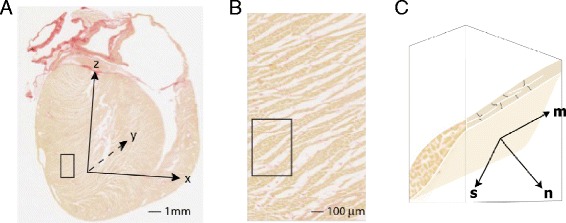
Figure 1.

Local structure-based coordinate system. A - long axis histological section from rat heart indicating the cardiac coordinate system (x,y,z). The z axis passes through the left ventricle apex and the center of the mitral valve orifice. B - magnified view of the region identified by box in (A). C - schematic 3D representation of a single layer of myocytes within the sub-region indicated in (B). This representation is simplified to facilitate labelling of the local structure-based coordinate system. The lamina consists of branching myocytes and is bounded by a network of perimysial collagen. In the local axis system, m aligns with the myocyte axis, n is normal to surface of the lamina and s is orthogonal to m and n. This cartoon does not show important microstructural features. These include i) branching and interconnection of laminae ii) curvature of laminae and myocyte orientation, and iii) the existence of adjacent laminae with different orientations.
