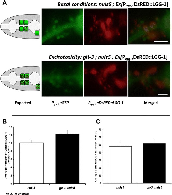
Figure 3.

A DsRed::LGG-1 reporter of autophagy does not provide convincing evidence for triggering of autophagy in at-risk neurons exposed to the excitotoxic insult. A) Representative images showing at-risk neurons in green (expressing P glr-1 ::GFP) and DsRed::LGG-1 expressing cells in red. Lateral view, anterior left, dorsal up, illustration on the left describing the results expected from a putative involvement of autophagy in excitotoxicity. Expression of green labeling in the pharynx comes from the co-injection marker for the DsRed::LGG-1 label, expressing P myo-2 ::GFP. B) Analysis of images taken from the two groups shows a similar number of at-risk neurons (green cells) showing DsRed::LGG-1 puncta. The observed small difference is not statistically significant. (t test used here) C) The average intensity of the DsRed::LGG-1 signal in at-risk neurons (green) in very similar in the two groups.
