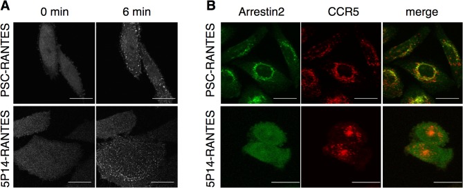
Fig 5. Spatial and temporal resolution of arrestin2-CCR5 association.
A CHO-CCR5 cells stably transfected with arrestin2-GFP were treated with chemokine analogs (100 nM) as indicated and the redistribution of arrestin2-GFP was followed by live fluorescence microscopy. Images captured prior to (0 min) and after ligand treatment (6 min) are shown. B CHO-CCR5 cells stably transfected with arrestin2-GFP (green), preincubated with rhodamine-labeled anti-CCR5 antibody (red), were washed and then incubated 90 min at 37°C with chemokine analogs (100 nM) prior to image capture. Maximal intensity projections are shown. Scale bar = 20 μm.

