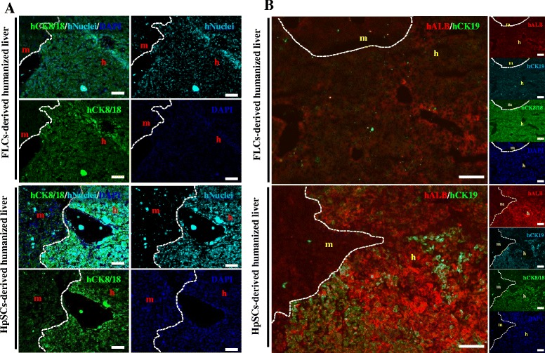
Figure 4.

Characterization of human primary fetal liver cell- and human hepatic stem cell-derived human hepatocytes in Alb-TRECK/SCID mice. (A) Immunohistochemistry to distinguish between human hepatocytes stained with anti-human CK8/18 (green) antigen and anti-human nuclear antigen (aqua blue) in human primary fetal liver cell- (FLC; upper panels) and human hepatic stem cell- (HpSC; lower panels) derived humanized livers at 6 weeks after transplantation. (B) Immunohistochemistry analyses for human albumin, human CK19, and human CK8/18 expression in human primary FLC- (upper panels) and HpSC- (lower panels) derived livers at 6 weeks after transplantation. White dashed line: mouse liver region distinguished from human liver region. Nuclei were counterstained with 4′,6-diamidino-2-phenylindole (DAPI, blue). Scale bars = 100 μm. m, mouse liver region; h, human donor cell-derived human region.
