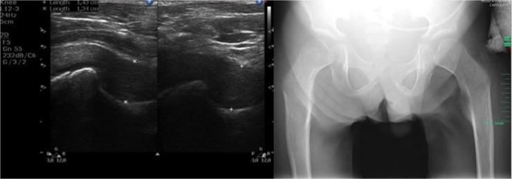
Fig 3. Ultrasound and X-ray images of hip joints.

(Left) Transverse scan of hip joint a 12-year-old patient with a severe phenotype of MPS II. (Right) Radiograph of the pelvis of a 12-year-old patient with a severe phenotype of MPS II: dysostosis multiplex (irregular shape of the pelvis, hypoplastic hip acetabulum, dysplastic hips, osteonecrosis of the femoral heads with flattened acetabula, lopsided head of femur bones).
