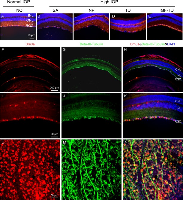
Fig 4. RGC loss in the experimental glaucoma model.
(A—E) Retina sections immmunostained for ß-III tubulin (red fluorescence) confirm microbead-induced glaucomatous RGC loss (SA, NP and TD) and RGC rescue in eyes transplanted with hNPIGF-TD cells (IGF-TD). Brn3a antibody (red fluorescence) was used to verify the accuracy of RGC density stained with ß-III tubulin (green fluorescence) on retinal cross sections. (F—K) and retinal flatmounts using confocal microscopy (L—N). RGC population was slightly overestimated (1.7%) using ß-III tubulin staining compared to Brn3a staining, but the difference was not significant (Mann-Whitney U test, P > 0.05). Abbreviations: ONL, outer nuclear layer; INL, inner nuclear layer; RGC, retinal ganglion cell; IGF-TD, transplanted hNPIGF-TD cells after microbead injection; TD, transplanted hNPTD cells after microbead injection. hNP, untransfected hNPs after microbead injection; SA, intravitreal saline (no cells) injection after microbead injection; NO, intravitreal saline injection and saline injection into the anterior chamber (no microbead and cell injection). High IOP, elevated intraocular pressure by microbead injection. Scale bar: 50 μm in A—E.

