
Figure 10.
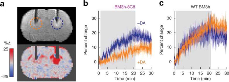
Detecting a metabolite with a responsive MRI contrast agent. The BM3h-8C8 agent was developed through directed evolution to bind dopamine to the protein’s heme group. a) A coronal MR image from a rat injected with BM3h-8C8 in the presence (orange dashed circle) or absence (blue dashed circle) of equimolar dopamine. MRI hyperintensity is noticeable near the tip of the dopamine-free cannula, indicating a short T1 relaxation time constant, while the lack of hyperintensity at the dopamine cannula indicates a long T1 relaxation time constant for the system in the presence of this agent. b,c) The temporal change in T1-weighted MRI signal shows a relative decrease in T1-weighting in the presence of dopamine (orange) in the rat treated with BM3h-8C8 relative to the rat treated with the wildtype BM3h protein that does not bind to dopamine. Reproduced with permission from reference (121).