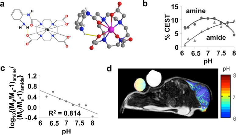
Figure 13.

A MRI contrast agent that measures extracellular pH within in vivo tumor tissue. a) 2-dimensional and 3-dimensional models of Yb-DO3A-oAA shows that the proximity of Yb(III) to the amide and the amine causes a shift in the magnetic resonance frequencies of these labile protons, which facilitates CEST MRI studies. b) The CEST effects from the amide and amine are dependent on pH. c) A log10 ratio of the CEST effects is linearly related to pH. d) A parametric pH map of a mouse tumor model overlaid on an anatomic MR image shows an acidic extracellular environment in the tumor region. The agent was directly injected into the tumor tissue to generate strong CEST signals in the tumor. Significant CEST signals were not detected elsewhere in the mouse model. Reproduced with permission from (174).
