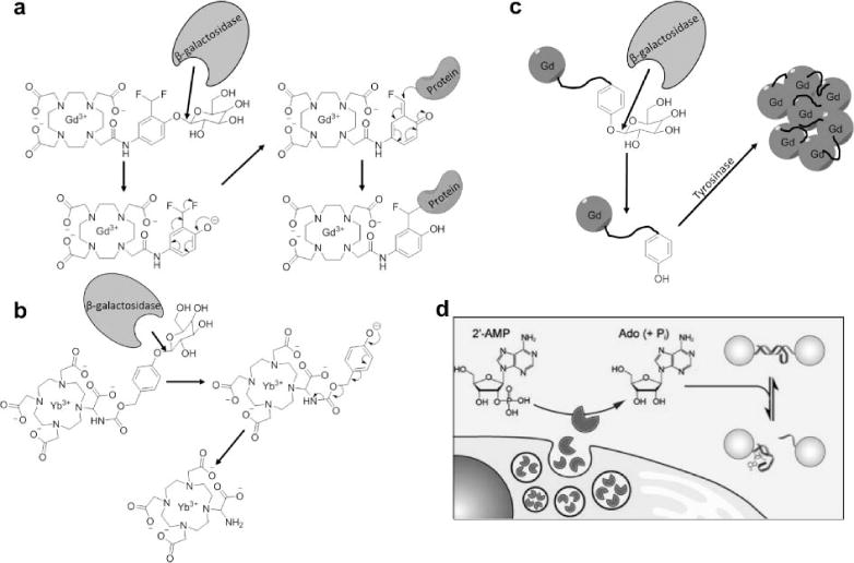
Figure 7.

Responsive MRI contrast agents can detect enzyme activities using a multistep approach. a) β-galactosidase hydrolyzes a β-galactopyranose ligand, forming a reactive phenolate anion that binds the contrast agent to a protein, slowing the tumbling time of the contrast agent and decreasing T1 relaxation time constant (59). b) β-galactosidase hydrolyzes the same ligand on CEST agent, forming an electron donating group that promotes aromatic delocalization that creates an amine group that can generate CEST (55). c) β-galactosidase-catalyzed hydrolysis of the same ligand creates a tyrosine ligand that can be polymerized by tyrosinase, creating a large molecular system with a slower tumbling time with decreased T1 relaxation time constant (62). d) Secreted alkaline phosphatase de-phosphorylates 2′-AMP to create adenosine, which can disrupt a DNA duplex that links SPIONs, which increases T2* relaxation time constant. Figure 7d was reproduced with permission from (80).
