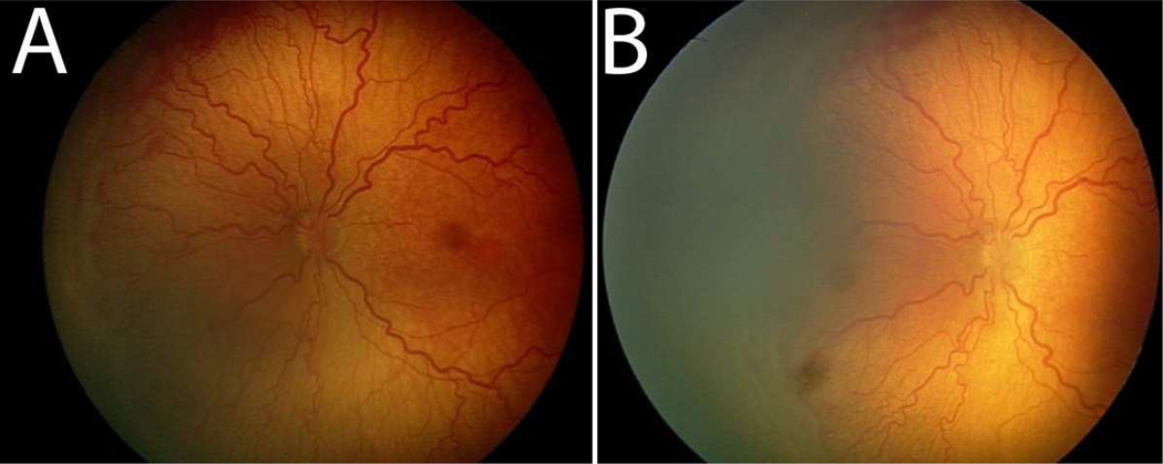
Figure 1.
Pre- and post-treatment images of the retina after intravitreal bevacizumab. Prior to treatment, plus disease with prominent tortuosity and congestion of the retinal vessels are observed in the left eye of a patient with Type 1 ROP (A). Two weeks post-treatment, the retinal vesels appear significantly less tortuous and congested (B).

