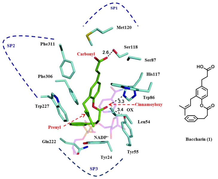
Figure 2.
Model of the AKR1C3.NADP+.Baccharin complex (left) and structure of baccharin in the orientation found in the model (right). AKR1C3 residues (light blue); NADP+ (pink); and baccharin (green). Dotted line: possible hydrogen bond; OX: oxyanion site; SP: subpocket. The docking model of the AKR1C3. NADP+.Baccharin complex was constructed by conducting calculations using the program Glide 5.0. The generated docked structure showing the highest score was selected for further structural analysis. The crystal structure of AKR1C3.NADP+ complex was chosen from RCSB protein data bank (PDB code: 1S2C) as a starting template. Reprinted with permission from (S. Endo et al., Selective inhibition of human type-5 17β-hydroxysteroid dehydrogenase (AKR1C3) by baccharin, a component of Brazilian propolis, Journal of Natural Products [37]). Copyright 2012, American Chemical Society.

