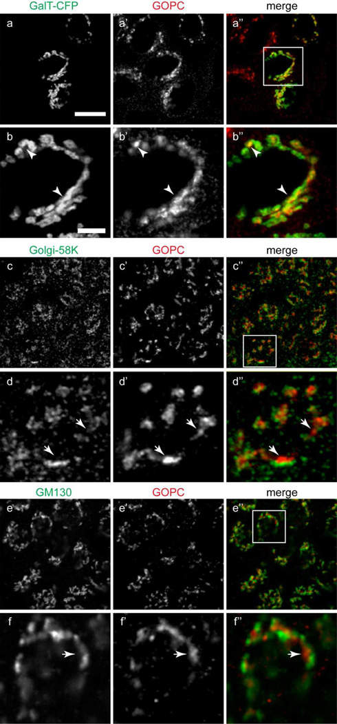
Figure 2.
(a–b”) MDCK cells transfected with the TGN-marker enzyme galactosyltransferase-CFP (a), fixed and labeled for GOPC (a’). b-b” show the inset region in image a”. There is extensive overlap of the markers (b-b”, arrowheads). (c–d”) Cells double-labeled with antibodies against the trans-Golgi cisternae marker Golgi-58K (c) and GOPC (c’). d-d” show the inset area in image c”. The labeling is in adjacent cisternae (d-d”, arrows). (e–f”) Cells double-labeled with antibodies against the medial Golgi marker GM130 (e) and GOPC (e’). f-f” show the inset area in image e”. The merged image shows no overlap of the markers (f-f”, arrows). Scale bar top panel, 20µm, bottom panel, 1µm. Representative images are from at least 4 experiments.

