Figure 6.
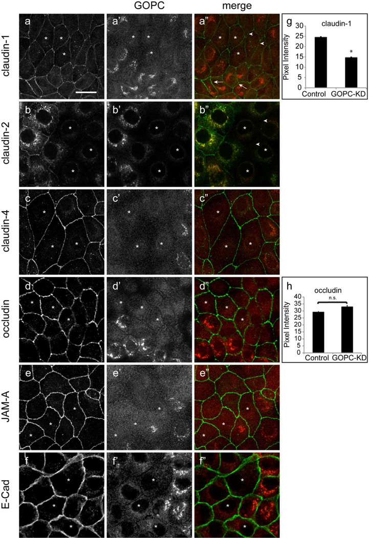
Knockdown of GOPC results in reduced claudin-1 at the tight junction. GOPC knockdown cells were double labeled for GOPC (a’, b’, c’, d’, e’, f’) and claudin-1 (a), claudin-2 (b), claudin-4(c), occludin (d), JAM-A (e), and E-cadherin (f). Knockdown of GOPC results in decreased claudin-1 at the lateral membrane (a”, arrowheads). Normal lateral membrane claudin-1 labeling is observed in adjacent cells that express GOPC (a”, arrows). Claudin-2 is also absent from cells knocked down for GOPC (b”, arrowheads). However, claudin-4, occludin, JAM-A, and E-cadherin labeling is unaffected by GOPC knockdown (c”, d”, e”, f”). Knockdown cells are indicated with (*). Scale bar, 20µm. (g,h) NIH-ImageJ quantification of pixel intensity of claudin-1 (n=52) (g) and occludin (n=37) labeling (h) at the lateral membrane after GOPC knockdown. *p < 0.01.

