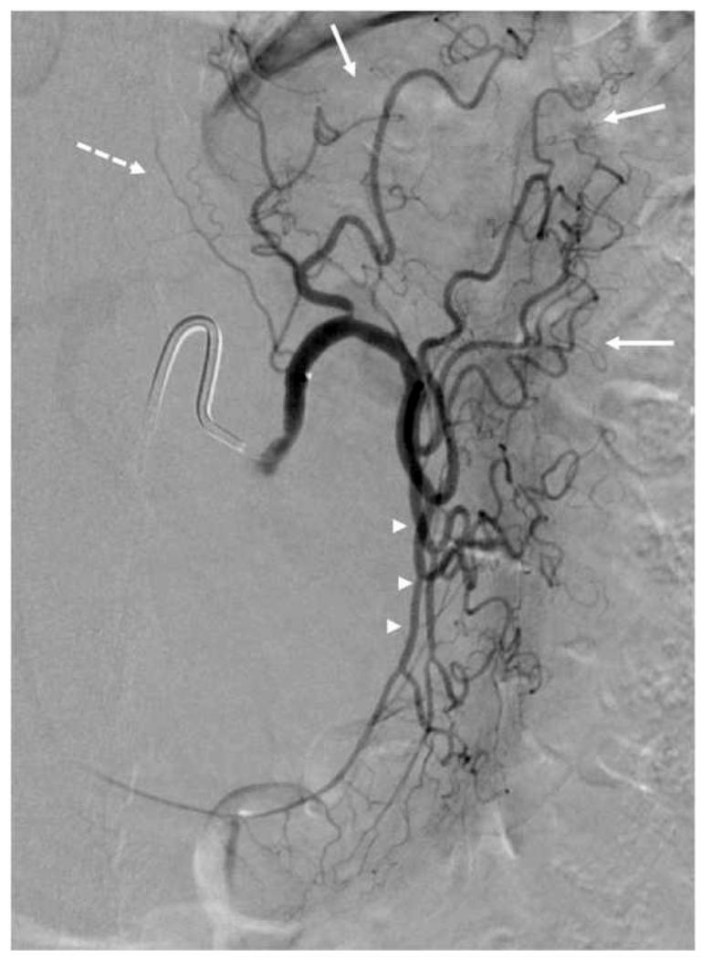
Figure 6. Digital Subtraction Angiogram (DSA) of the Left Gastric Artery.
Left gastric artery selected using a 5F SOS-selective catheter and a high-flow micro-catheter. Fundal (solid arrow) and esophageal (dashed arrow) branches are identified. This left gastric artery demonstrates a large anastomosis (arrowheads) with the right gastric artery along the lesser curvature.
©Johns Hopkins University

