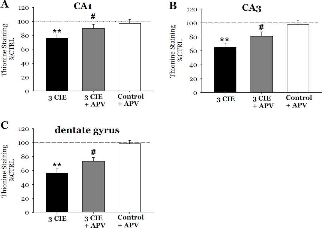
Figure 7.
Effects of exposure to 50 mM ethanol for 5 DIV, followed by exposure to APV (40µM) during the 24-hour period of ethanol withdrawal, and repeated for 3 CIE on thionine staining in organotypic slice cultures. Exposure to 50 mM ethanol for 5 DIV, followed by a 24-hour period of ethanol withdrawal, and repeated for 3 CIE resulted in consistent and significant decreases of thionine staining as compared to control values in the pyramidal cell layers of the CA1 (A) and CA3 (B), and granule cell layer of the dentate gyrus (C). Exposure to APV (40 µM) during periods of withdrawal attenuated the decreases of thionine staining in the CA1, CA3, and dentate gyrus; whereas exposure to APV in ethanol naïve slices did not significantly alter levels of thionine (Figure 7). **p <0.001 vs control; #p <0.05 vs 3 CIE.

