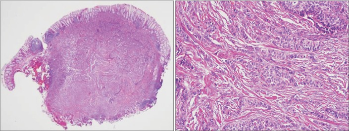
Fig. 2. Histopathological findings of the rectal neuroendocrine tumor. (A) The endoscopically resected specimen mostly consisted of a relatively well-demarcated tumor. The tumor involved the mucosa and submucosa, and it measured 8 mm across the greatest dimension. The deep resection margin characterized the tumor (H&E, ×10). (B) The tumor cells formed nests or cords in the sclerotic stroma and demonstrated histological patterns typical of a neuroendocrine tumor. The tumor cell nuclei are round or ovoid and demonstrate fine salt-and-pepper chromatin (H&E, ×200).

