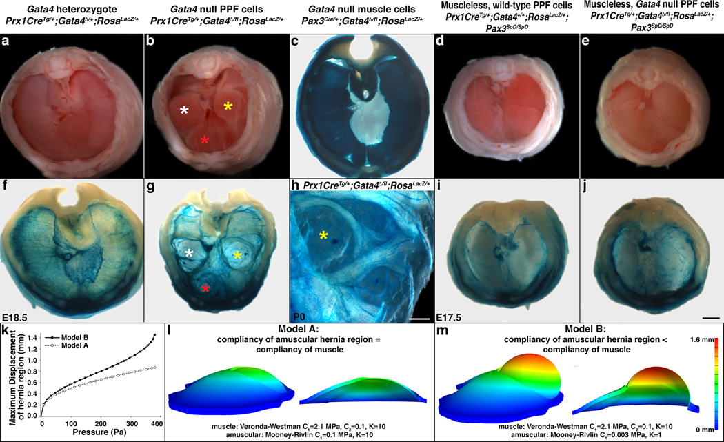
Fig. 2. Deletion of Gata4 in the PPFs produces localized amuscular regions that are weaker than juxtaposed muscular regions and results in CDH.
(a–d) CDH develops in mice with Gata4 null PPF cells (n > 33/33; Bochdalek hernias labeled with white and yellow asterisks and Morgagni hernia labeled with red asterisk in b), but not in Gata4 heterozygotes (n > 66/66; a), mice with Gata4 null muscle (n = 28/28; c), or mice with muscleless diaphragms (n > 10/10; d). (e, j) Loss of muscle in diaphragms with Gata4 null PPF cells rescues herniation, indicating that juxtaposition of amuscular with muscular regions is required for CDH (n = 3/3). (g–h) In herniated regions PPF cells are present, but muscle is not (n = 7/7). (k–m), Finite element modeling shows that a hernia only develops when the amuscular region is more compliant than the muscular region. Models are lateral views (c, f–j) Whole-mount β-galactosidase staining. (a–j) Dorsal is at top. Scale bar = 1 mm (a–g, i–j), 0.5 mm (h).

