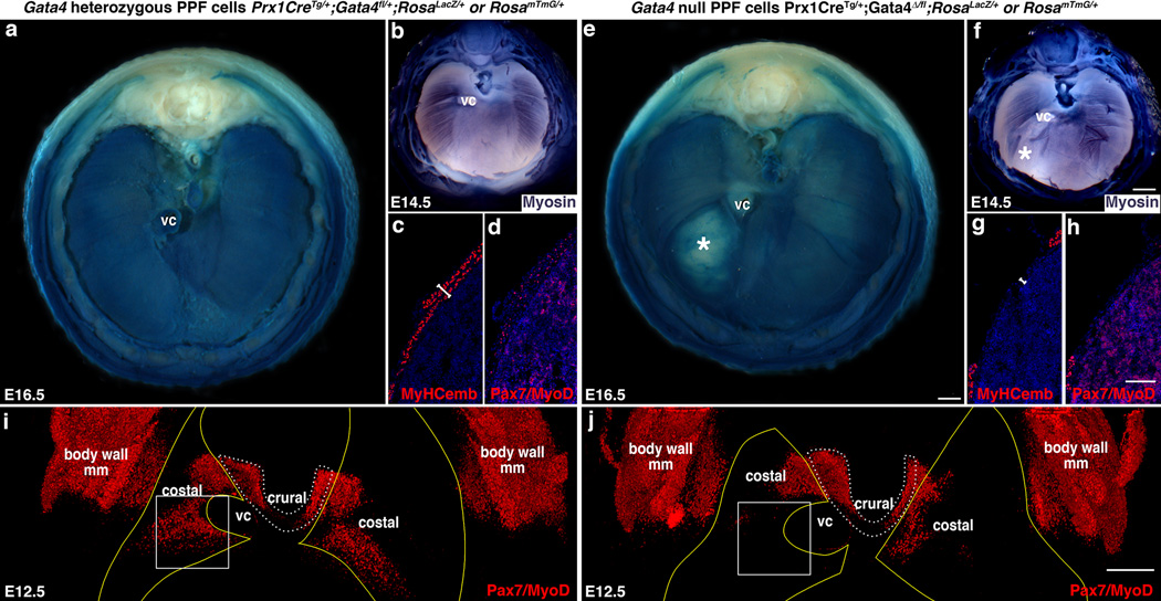
Fig. 4. CDH results from early defects in the localization of muscle progenitors.
(a, e) Overt liver herniation through diaphragms with Gata4 null PPF cells first appears at E16.5 (* in e, n = 3/3). (b–d, f–h) At E14.5 differentiating myofibers are aberrant (* in f; n = 7/7; b, f) and myofibers (c, g) and Pax7+/MyoD+ muscle progenitors (d, h) are absent in localized regions (n = 1/1). (i–j) At E12.5 Pax7+/MyoD+ muscle progenitors are absent in localized regions, particularly in the region (box) that consistently gives rise to hernias (n = 7/7). Pleuroperitoneal folds are outlined in yellow (outline derived from GFP immunofluorescence shown in Fig. 6a and b) and costal muscle is outlined in white dashed lines. (a, b) Whole-mount β-galactosidase staining. (b, f) Whole-mount myosin-alkaline phosphatase staining. (c–d, g-–h) Section immunofluorescence. (i-j) Whole-mount immunofluorescence (same diaphragms shown in Fig. 6a and b). (a–b, e–f, i–j) Dorsal is at the top. Scale bar = 500 µm (a, e), 500 µm (b, f), 100 µm (c–d, g–h), 200 µm (i–j). VC, vena cava; body wall mm, body wall muscles.

