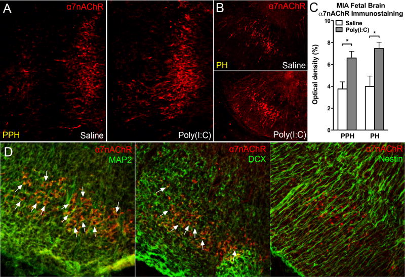
Fig. 6.
MIA increased α7nAChR in fetal brain neurons. MIA E12.5 fetal hindbrain displayed more α7nAChR immunostaining than control hindbrain 3 hours following maternal poly(I:C) injection. In hindbrain subregions (A) PPH and (B) PH, α7nAChR was increased in MIA offspring. (C) Optical density quantitation revealed the increased α7nAChR immunostaining in MIA fetal hindbrain. PPH: Saline: n = 3 litters, Poly(I:C): n = 3 litters; PH: Saline: n = 3 litters, Poly(I:C): n = 4 litters. (D) Confocal images demonstrated α7nAChR-positive cells were double-labeled with mature neuronal marker MAP2 (left panel) and the immature neuronal marker DCX (middle panel), but not with the neural stem cell marker nestin (right panel). White arrows indicate the colocalization between α7nAChR and the other markers. PPH: prepontine hindbrain, PH: pontine hindbrain, MAP2: microtubule-Associated Protein 2, DCX: doublecortin. Data are presented as mean ± SEM. Significant difference between groups is labeled as * p < 0.05.

