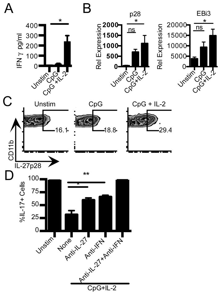
Figure 3. IL-2 promotes IFN-γ and IL-27 production from CD11b+ CD11c− APC.
A) CD11b+CD11c− cells from NOD were stimulated with CpG, or CpG+ IL-2. IFN-γ production in culture supernatant at 24 h (n=5) was determined by CBA. *p=0.0074, t-test. B) CD11b+CD11c− cells were stimulated as in (A) for 4 h. Cells were then lysed for RNA isolation and use in real-time PCR. Data are an average of 2–3 independent experiments. Left panel, IL-27 p28. Right panel, EBi3. *p<0.05, One-Way ANOVA, Tukey’s multiple comparison test. C) NOD splenocytes were T-depleted and stimulated for 18h with media (unstim), CpG, or CpG+IL-2. Staining for IL27 p28 in CD11b+CD11c− cells is shown. Data shown are representative of five independent experiments. D) Th17 differentiation of naïve NOD CD4+ T cells cultured with syngeneic T cell-depleted APC that were cultured for 18h with media (unstim), CpG+ IL-2 in the presence or absence of anti-IFN-γ and anti-IL-27 p28 as indicated. Data are normalized to frequency of Th17 cells in cultures with untreated APC. *p=0.0334,**p=0.0148, One-Way ANOVA, Tukey’s multiple comparison test.

