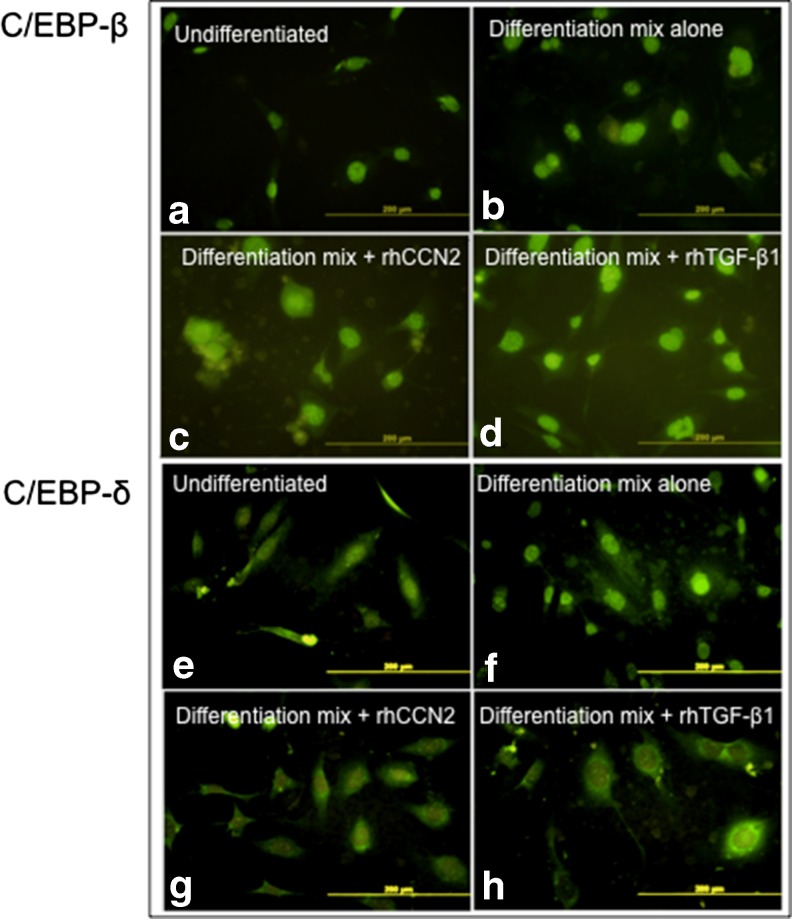
Fig. 2.
Nuclear compared with cytosolic localisation of C/EBP-β and C/EBP-δprotein by rhCCN2 or rhTGF-β1 each in the presence of differentiation mix. Representative immunoflourescence images of C/EBPs 24 h after addition of differentiation mix. Nuclear localisation of both C/EBP-β (a-d) and C/EBP-δ (e-h) are shown. NIH/3T3L1 cells were either non-differentiated (a, e) or they were treated with differentiation mix alone (b, f), or differentiation mix plus either added rhCCN2 (500 ng/ml) (c, g) or added active rhTGF-β1 (2 ng/ml) (d, h). Each size-bar indicates 200 μM

