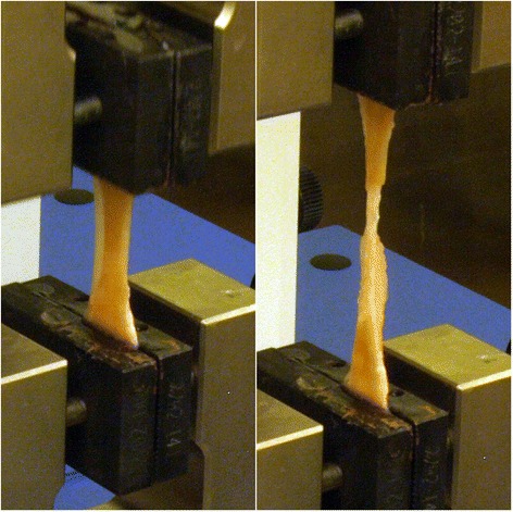
Figure 4.

Representative images of tissue deformation during tensile testing. Tissue was placed between two serrated grips, with an initial gauge length of 2 cm, and pulled at a constant rate of 1%/sec (Left). All tissue samples tested thinned mid-substance before ultimately failing through progressive tearing at the same location (Right).
