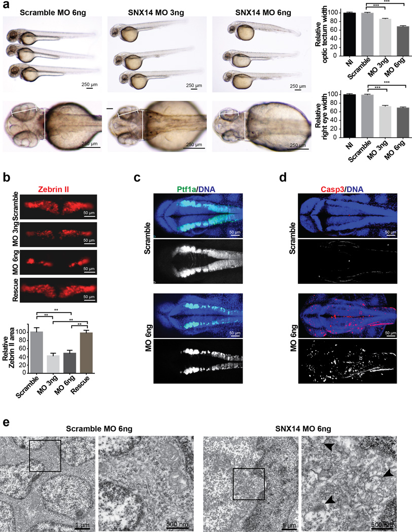
Figure 5. Morphant snx14 zebrafish show apoptosis, excessive autophagic vesicles, and loss of neural tissue including cerebellar primordium.
(a) Comparison of scrambled (6ng) and snx14 (3ng and 6ng) morphant zebrafish 48 hours postfertilization (hpf). Calipers: measured distance. Scale bar 250 µm. Graphs: Reduced optic tectum and right eye width in morphants. Mean ± SEM (n = 15 embryos for NI, 16 for Scramble, 31 for MO 3ng and 18 for MO 6ng, N = 2). *p < 0.05; **p < 0.005 (two tiled t-test). (b) Scramble or snx14 morphants for Zebrin II (Purkinje cell marker), rescued with human SNX14 (50 pg). Scale bar 50 µm. Graph: Zebrin II compartment area relative to scramble MO injected embryos. Mean ±SEM (n = 10 embryos for Scramble, 6 for MO 3ng, 9 for MO 6ng and 9 for rescue) *p < 0.05; **p < 0.005 (two tiled t-test). (c) Maximum confocal projection from 36 hpf Tg(ptf1a:eGFP) (green) zebrafish with scramble or snx14 MO showing reduced Purkinje cell progenitors. (d) Maximum confocal projection with increased caspase 3 (red) positive cells in 36 hpf snx14 morphants. Blue: DAPI. Scale bar 50 µm. (e) Transmission electron microscopy showing autophagic structures in 48 hpf snx14 and scrambled morphant neurons residing between the optic lobes. Box: Highlighted areas. Arrowheads: autophagic structures.

